Sir James Paget, MD lived in the 1800s. He is famous for describing two unrelated medical conditions that bear his name: Paget’s disease of the bone and Paget’s disease of nipple. This summary is about Paget’s disease of the nipple.
Paget’s disease of the nipple (sometimes referred to as Paget’s disease of the breast, it’s the same thing) is an uncommon form of breast cancer. I see about one or two cases a year. The theory in Paget’s disease of the nipple is that cells that line the milk ducts in the breast (called ductal cells) somehow become malignant and start travelling up the breast ducts to the ducts in the nipple and then go on to spread to the skin that overlies the nipple. So, if a patient has Paget’s of the nipple, it is assumed that there is probably also cancer deeper inside the breast.
The trick with Paget’s disease is to recognize it.
SIGNS & SYMPTOMS
Initially, it’s not unusual for Paget’s to either be ignored altogether or misdiagnosed as eczema, dermatitis, dry skin or infection. Patients often try topical antibiotics, anti-inflammatory ointments, steroid creams or even oral antibiotics but because Paget’s disease is not an infection the nipple does not get better. In these cases, the diagnosis becomes clearer over time by observing that the nipple skin does not respond to treatment. Failure to make the diagnosis immediately is not the end of the world because, fortunately, Paget’s is characteristically a slow-growing cancer. Since the visual appearance of Paget’s varies and can look different from one person to the next, my rule of thumb is, “If the nipple looks unusual, think Paget’s”.
The hallmark of Paget’s is dry, flaky or scaly skin on the nipple. No matter how much the patient tries to wash or peel away the dry skin and applies lotions, creams, etc., the dry, flaky skin keeps coming back. Over the years, it has been my observation that Paget’s appears on the nipple first and then extends to the areola and eventually progresses onto the skin of the breast.
Usually, the nipple is pink or red and can look a little raw. There may be some clear or yellow serous fluid and when it dries, the serous fluid becomes crusty. Technically this is not nipple discharge, which is where fluid comes from the milk ducts. Rather the serous fluid and crusting in Paget’s is due to loss of the epithelial skin layer and subsequent weeping of serous fluid from the raw dermis. In addition to dryness, flakiness and peeling, there is skin thickening and sometimes the nipple changes shape and may become flat or inverted.
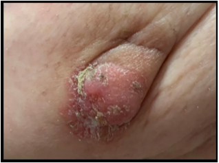
This is a photo of one of my patients who had Paget’s disease of the nipple. Note the dry, flaking superficial skin and the underlying skin which is pink and thickened. There is obvious crusting. After a trial of topical steroids, antifungals and oral antibiotics, her primary doctor referred the patient to me. The unsuccessful trial of medications made my job easy. I went straight to a full thickness biopsy which showed Paget’s disease of the nipple.
The hallmark of Paget’s is a dry, flaky, scaly nipple.
Paget’s disease of the nipple is painless. The nipple does not hurt and it is not tender. However, it is not uncommon for the nipple to be itchy in Paget’s disease. Having said that, the opposite is not true. In other words, not all itchy nipples are due to Paget’s disease. The sensation of itchiness in one or both nipples is quite common and without visible changes in the skin, an itchy nipple alone is not a symptom of Paget’s disease. Theoretically both nipples can have Paget’s disease at the same time but I’ve only ever seen unilateral Paget’s. Finally, Paget’s does not seem to be related to breast feeding or nipple piercing.
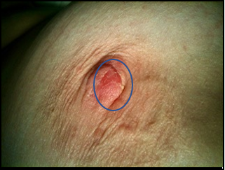
This patient was referred to me because her left mammogram was abnormal. When I examined her in the office, I noticed that a portion of the right nipple looked different than the left. The inner part was a little red, with thick skin and yellow crusting. So, I biopsied the abnormal area seen on the left mammogram and did a full-thickness biopsy of the right nipple. The left side was benign but the right nipple showed Paget’s disease.
DIAGNOSIS & TREATMENT
The only way to know for sure what is going on with an abnormal looking nipple is to do a biopsy. A full thickness piece of the nipple needs to be excised and examined under the microscope. As with all types of breast cancer, this is the only way to definitively make a diagnosis. You can’t just scrape off some of the dead skin. There is no blood test or anything specific on breast imaging. You need a piece of tissue. Sometimes the tissue is obtained by a dermatologist in the form of a punch biopsy. When I do a biopsy for suspected Paget’s, I remove a small wedge of the nipple. This is usually done under general anesthesia. The recovery time after a full thickness biopsy is 2 or 3 days and the cosmetic outcome afterwards is excellent.
If a patient has biopsy-proven Paget’s disease of the nipple, it is assumed that there is also cancer deeper inside the breast. We look for additional cancer using mammogram, ultrasound and sometimes MRI. Despite the assumption, there are times when we don’t find any additional cancer in the breast. In this circumstance, it’s believed that the nipple ducts became cancerous on their own. Paget’s disease of the nipple is classified as stage 0 breast cancer and since stage is related to prognosis, Paget’s disease on its own has a very good prognosis. Most of the time however, there is adjacent breast cancer and usually it is intraductal carcinoma or ductal carcinoma in situ (DCIS) which is also stage 0. Sometimes the adjacent breast cancer is invasive cancer.
The treatment of Paget’s is the same as with DCIS or invasive cancer (see Treatment), with one important addition: the pagetoid area must be surgically removed with negative margins. In my experience, this usually requires removal of the entire nipple areolar complex (NAC).