Scout
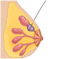
The Savi Scout is a relatively new surgical guidance system and is a good alternative to wire-guided localization.There are 3 components: the SCOUT reflector, the handpiece and the console.
The reflector is an infrared-activated device that is put into the breast by the radiologist with image guidance prior to surgery. It can be inserted at a facility other than where the surgery will be done. This is important because it eliminates the time-consuming work of tracking down and obtaining outside films and having them reviewed by the hospital radiologist. The Scout reflector is not radioactive, has no external components, and is FDA–approved for placement up to 30 days before the surgical procedure. It is 12-mm long and consists of an infrared light receptor, resistor, and two antennae. It is slightly larger than the tissue marker/clip placed during needle biopsy.
The handpiece and console are used by the surgeon in the operating room. The console sends a radar signal and infrared light to the disposable handpiece which then delivers the radar signal and infrared light into the breast and receives signals reflected back from the reflector. The console processes the reflected radar signal to provide the surgeon with reflector proximity and location information using audible and visual signals. The numeric display provides real-time distance between the handpiece and reflector. The audible signal increases in cadence when the handpiece is closer to the reflector. The maximum allowable distance from the handpiece to the reflector is 6 cm. The audible and visual signals guide the surgical dissection through the breast to the tumor.

Scout reflector
Once the targeted area is localized, it is excised using standard surgical technique. After the lesion has been removed, a specimen mammogram is obtained to confirm that the area of concern, the Scout reflector and the tissue marker/clip have all been removed. This is the same protocol as is done with wire-localization.
Advantages of Scout compared to wire-localization
– makes scheduling surgery easier
– shortens the time spent in hospital
– can be inserted at another location at a time convenient for the patient
Disadvantages of Scout compared to wire-localization
– Scout cost more
– patient has to make one extra visit
– cannot be used if the lesion is more than 6 cm away from the surface of the breast
No difference between Scout and wire-localization
– pain
– re-excision for positive margins
– amount of tissue removed
– complication rate